3M7R | pdb_00003m7r

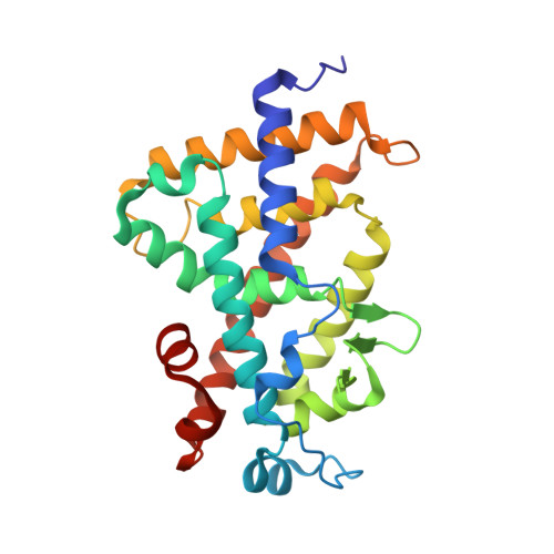
Crystal structure of VDR H305Q mutant


Experimental Data Snapshot

  • Method: X-RAY DIFFRACTION
  • Resolution: 1.80 ?
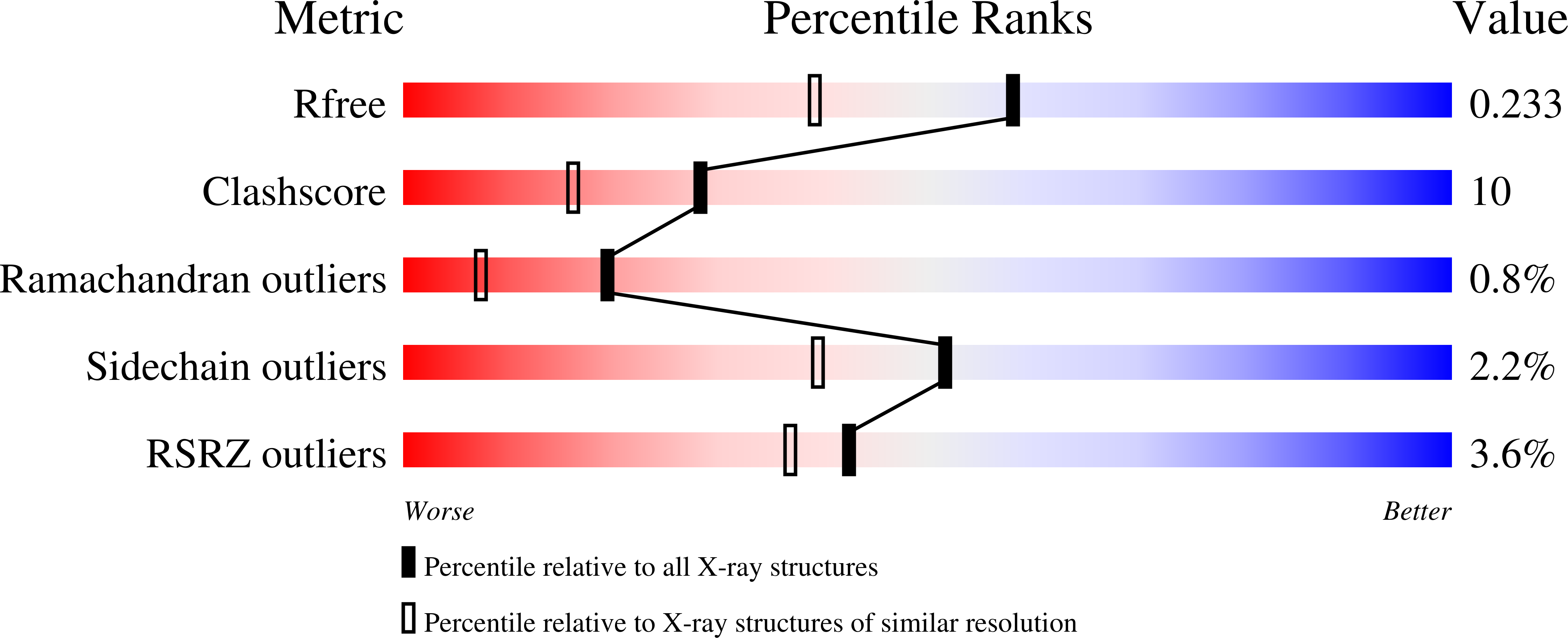
  • R-Value Free: 
    0.241 (Depositor), 0.230 (DCC) 
  • R-Value Work: 
    0.211 (Depositor), 0.200 (DCC) 
  • R-Value Observed: 
    0.241 (Depositor) 

Starting Model: experimental
View more details

wwPDB Validation   3D Report Full Report


Ligand Structure Quality Assessment 


This is version 1.5 of the entry. See complete history


Literature

Crystal structure of hereditary vitamin D-resistant rickets--associated mutant H305Q of vitamin D nuclear receptor bound to its natural ligand

Rochel, N.Hourai, S.Moras, D.

(2010) J Steroid Biochem Mol Biol 121: 84-87

  • DOI: https://doi.org/10.1016/j.jsbmb.2010.04.008
  • Primary Citation of Related Structures:  
    3M7R

  • PubMed Abstract: 

    In the nuclear receptor of vitamin D (VDR) histidine 305 participates to the anchoring of the ligand. The VDR H305Q mutation was identified in a patient who exhibited the hereditary vitamin D-resistant rickets (HVDRR). We report the crystal structure of human VDR H305Q-ligand binding domain bound to 1alpha,25(OH)2D3 solved at 1.8A resolution. The protein adopts the active conformation of the wild-type liganded VDR. A local conformational flexibility at the mutation site weakens the hydrogen bond between the 25-OH with Gln305, thus explaining the lower affinity of the mutant proteins for calcitriol. The structure provides the basis for a rational approach to the design of more potent ligands for the treatment of HVDRR.


  • Organizational Affiliation

    IGBMC (Institut de G¨¦n¨¦tique et de Biologie Mol¨¦culaire et Cellulaire), D¨¦partement de Biologie et de G¨¦nomique Structurales, Centre National de la Recherche Scientifique, Institut National de la Sant¨¦ de la Recherche M¨¦dicale, Universit¨¦ de Strasbourg, 1 rue Laurent Fries, Illkirch, France. rochel@igbmc.fr


Macromolecules
Find similar proteins by:  (by identity cutoff)  |  3D Structure
Entity ID: 1
MoleculeChains Sequence LengthOrganismDetailsImage
Vitamin D3 receptor253Homo sapiensMutation(s): 1 
Gene Names: NR1I1VDR
UniProt & NIH Common Fund Data Resources
Find proteins for P11473 (Homo sapiens)
Explore P11473 
Go to UniProtKB:  P11473
PHAROS:  P11473
GTEx:  ENSG00000111424 
Entity Groups  
Sequence Clusters30% Identity50% Identity70% Identity90% Identity95% Identity100% Identity
UniProt GroupP11473
Sequence Annotations
Expand
  • Reference Sequence
Small Molecules
Ligands 1 Unique
IDChains Name / Formula / InChI Key2D Diagram3D Interactions
VDX
Query on VDX

Download Ideal Coordinates CCD File 
B [auth A]5-{2-[1-(5-HYDROXY-1,5-DIMETHYL-HEXYL)-7A-METHYL-OCTAHYDRO-INDEN-4-YLIDENE]-ETHYLIDENE}-4-METHYLENE-CYCLOHEXANE-1,3-DIOL
C27 H44 O3
GMRQFYUYWCNGIN-NKMMMXOESA-N
Experimental Data & Validation

Experimental Data

  • Method: X-RAY DIFFRACTION
  • Resolution: 1.80 ?
  • R-Value Free:  0.241 (Depositor), 0.230 (DCC) 
  • R-Value Work:  0.211 (Depositor), 0.200 (DCC) 
  • R-Value Observed: 0.241 (Depositor) 
Space Group: P 21 21 21
Unit Cell:
Length ( ? )Angle ( ? )
a = 44.402¦Á = 90
b = 51.67¦Â = 90
c = 131.888¦Ã = 90
Software Package:
Software NamePurpose
CNSrefinement
HKL-2000data reduction
HKL-2000data scaling
CNSphasing

Structure Validation

View Full Validation Report


Ligand Structure Quality Assessment 


Entry History 

Deposition Data

Revision History  (Full details and data files)

  • Version 1.0: 2010-05-05
    Type: Initial release
  • Version 1.1: 2011-07-13
    Changes: Version format compliance
  • Version 1.2: 2014-02-19
    Changes: Database references
  • Version 1.3: 2018-02-07
    Changes: Advisory, Experimental preparation
  • Version 1.4: 2021-11-10
    Changes: Advisory, Database references, Derived calculations
  • Version 1.5: 2023-11-01
    Changes: Data collection, Refinement description