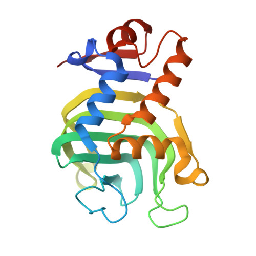
Replacing the Axial Ligand Tyrosine 75 or Its Hydrogen Bond Partner Histidine 83 Minimally Affects Hemin Acquisition by the Hemophore HasAp from Pseudomonas aeruginosa.
Kumar, R., Matsumura, H., Lovell, S., Yao, H., Rodriguez, J.C., Battaile, K.P., Moenne-Loccoz, P., Rivera, M.(2014) Biochemistry 53: 2112-2125
- PubMed: 24625274
- DOI: https://doi.org/10.1021/bi500030p
- Primary Citation of Related Structures:
4O6Q, 4O6S, 4O6T, 4O6U - PubMed Abstract:
Hemophores from Pseudomonas aeruginosa (HasAp), Serratia marcescens (HasAsm), and Yersinia pestis (HasAyp) bind hemin between two loops. One of the loops harbors conserved axial ligand Tyr75 (Y75 loop) in all three structures, whereas the second loop (H32 loop) contains axial ligand His32 in HasAp and HasAsm, but a noncoordinating Gln32 in HasAyp. Binding of hemin to the Y75 loop of HasAp or HasAsm causes a large rearrangement of the H32 loop that allows His32 coordination. The Q32 loop in apo-HasAyp is already in the closed conformation, such that binding of hemin to the conserved Y75 loop occurs with minimal structural rearrangement and without coordinative interaction with the Q32 loop. In this study, structural and spectroscopic investigations of the hemophore HasAp were conducted to probe (i) the role of the conserved Tyr75 loop in hemin binding and (ii) the proposed requirement of the His83-Tyr75 hydrogen bond to allow the coordination of hemin by Tyr75. High-resolution crystal structures of H83A holo-HasAp obtained at pH 6.5 (0.89 ?) and pH 5.4 (1.25 ?) show that Tyr75 remains coordinated to the heme iron, and that a water molecule can substitute for N¦Ä of His83 to interact with the O¦Ç atom of Tyr75, likely stabilizing the Tyr75-Fe interaction. Nuclear magnetic resonance spectroscopy revealed that in apo-Y75A and apo-H83A HasAp, the Y75 loop is disordered, and that disorder propagates to nearby elements of secondary structure, suggesting that His83 N¦Ä-Tyr75 O¦Ç interaction is important to the organization of the Y75 loop in apo-HasA. Kinetic analysis of hemin loading conducted via stopped-flow UV-vis and rapid-freeze-quench resonance Raman shows that both mutants load hemin with biphasic kinetic parameters that are not significantly dissimilar from those previously observed for wild-type HasAp. When the structural and kinetic data are taken together, a tentative model emerges, which suggests that HasA hemophores utilize hydrophobic, ¦Ð-¦Ð stacking, and van der Waals interactions to load hemin efficiently, while axial ligation likely functions to slow hemin release, thus allowing the hemophore to meet the challenge of capturing hemin under inhospitable conditions and delivering it selectively to its cognate receptor.
Organizational Affiliation:
Department of Chemistry, University of Kansas , Multidisciplinary Research Building, 2030 Becker Drive, Lawrence, Kansas 66047, United States.


