Structures of Hsp90 alpha and Hsp90 beta bound to a purine-scaffold inhibitor reveal an exploitable residue for drug selectivity.
Huck, J.D., Que, N.L.S., Sharma, S., Taldone, T., Chiosis, G., Gewirth, D.T.(2019) Proteins 87: 869-877
- PubMed: 31141217
- DOI: https://doi.org/10.1002/prot.25750
- Primary Citation of Related Structures:
6N8W, 6N8X, 6N8Y, 6OLX - PubMed Abstract:
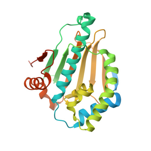
Hsp90¦Á and Hsp90¦Â are implicated in a number of cancers and neurodegenerative disorders but the lack of selective pharmacological probes confounds efforts to identify their individual roles. Here, we analyzed the binding of an Hsp90¦Á-selective PU compound, PU-11-trans, to the two cytosolic paralogs. We determined the co-crystal structures of Hsp90¦Á and Hsp90¦Â bound to PU-11-trans, as well as the structure of the apo Hsp90¦Â NTD. The two inhibitor-bound structures reveal that Ser52, a nonconserved residue in the ATP binding pocket in Hsp90¦Á, provides additional stability to PU-11-trans through a water-mediated hydrogen-bonding network. Mutation of Ser52 to alanine, as found in Hsp90¦Â, alters the dissociation constant of Hsp90¦Á for PU-11-trans to match that of Hsp90¦Â. Our results provide a structural explanation for the binding preference of PU inhibitors for Hsp90¦Á and demonstrate that the single nonconserved residue in the ATP-binding pocket may be exploited for ¦Á/¦Â selectivity.
Organizational Affiliation:
Hauptman-Woodward Medical Research Institute, Buffalo, New York.


