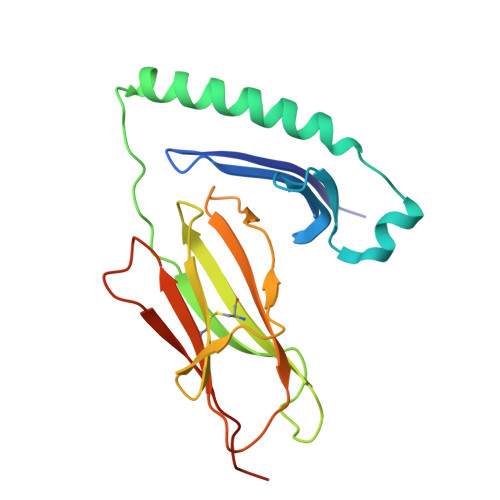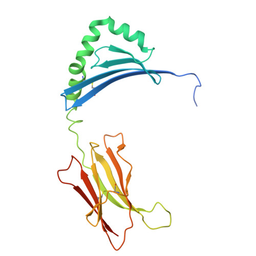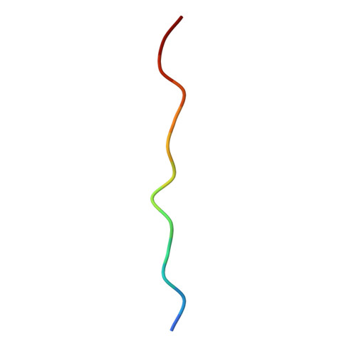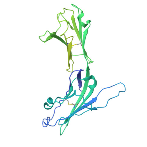
Crystal structure of the human LAG-3-HLA-DR1-peptide complex.
Petersen, J., Llerena, C., Golzarroshan, B., Faoro, C., Triebel, F., Rossjohn, J.(2024) Sci Immunol 9: eads5122-eads5122
- PubMed: 39671469
- DOI: https://doi.org/10.1126/sciimmunol.ads5122
- Primary Citation of Related Structures:
9BF9 - PubMed Abstract:
T cell activity is governed by T cell receptor (TCR) signaling and constrained by immune checkpoint molecules, including programmed cell death protein 1 (PD-1), cytotoxic T lymphocyte-associated antigen 4 (CTLA-4), and lymphocyte activation gene 3 (LAG-3). The basis for how LAG-3 binds to human leukocyte antigen class II molecules (HLA-II) remains unknown. Here, we present the 3.4-angstrom crystal structure of a LAG-3-peptide-HLA-II complex and probe the energetics of the complex interface. Coincident with the HLA-II binding site of the ancestrally related, monomeric CD4 receptor, the LAG-3 homodimer laterally engages two HLA-II molecules via distal D1 domain surfaces, imposing a 38° angular offset. The LAG-3-HLA-II interface is discontinuous and lacks involvement of the D1 extra loop, a binding site for anti-LAG-3 therapeutic monoclonal antibodies. Upon HLA-II binding, intrinsically mobile loops of the LAG-3 molecule become ordered, with contact residues highly conserved across HLA-DR, DQ, and DP allomorphs. Our data provide a structural foundation for development of immunomodulatory approaches targeting LAG-3.
Organizational Affiliation:
Infection and Immunity Program and Department of Biochemistry and Molecular Biology, Biomedicine Discovery Institute, Monash University Clayton, Victoria, Australia.