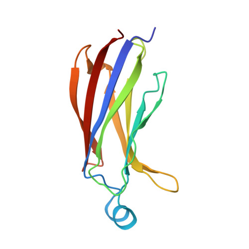
Structural basis of phosphatidylcholine recognition by the C2-domain of cytosolic phospholipase A2alpha.
Hirano, Y., Gao, Y.G., Stephenson, D.J., Vu, N.T., Malinina, L., Simanshu, D.K., Chalfant, C.E., Patel, D.J., Brown, R.E.(2019) Elife 8
- PubMed: 31050338
- DOI: https://doi.org/10.7554/eLife.44760
- Primary Citation of Related Structures:
6IEJ - PubMed Abstract:
Ca 2+ -stimulated translocation of cytosolic phospholipase A 2 ¦Á (cPLA 2 ¦Á) to the Golgi induces arachidonic acid production, the rate-limiting step in pro-inflammatory eicosanoid synthesis. Structural insights into the cPLA 2 ¦Á preference for phosphatidylcholine (PC)-enriched membranes have remained elusive. Here, we report the structure of the cPLA 2 ¦Á C2-domain (at 2.2 ? resolution), which contains bound 1,2-dihexanoyl- sn -glycero-3-phosphocholine (DHPC) and Ca 2+ ions. Two Ca 2+ are complexed at previously reported locations in the lipid-free C2-domain. One of these Ca 2+ ions, along with a third Ca 2+ , bridges the C2-domain to the DHPC phosphate group, which also interacts with Asn65. Tyr96 plays a key role in lipid headgroup recognition via cation-¦Ð interaction with the PC trimethylammonium group. Mutagenesis analyses confirm that Tyr96 and Asn65 function in PC binding selectivity by the C2-domain and in the regulation of cPLA 2 ¦Á activity. The DHPC-binding mode of the cPLA 2 ¦Á C2-domain, which differs from phosphatidylserine or phosphatidylinositol 4,5-bisphosphate binding by other C2-domains, expands and deepens knowledge of the lipid-binding mechanisms mediated by C2-domains.
Organizational Affiliation:
Structural Biology Program, Memorial Sloan-Kettering Cancer Center, New York, United States.


