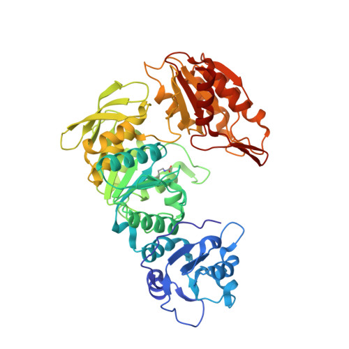
The crystal structure of Mycobacterium thermoresistibile MurE ligase reveals the binding mode of the substrate m-diaminopimelate.
Rossini, N.O., Silva, C., Dias, M.V.B.(2023) J Struct Biol 215: 107957-107957
- PubMed: 36944394
- DOI: https://doi.org/10.1016/j.jsb.2023.107957
- Primary Citation of Related Structures:
8G6P - PubMed Abstract:
The cytoplasmatic biosynthesis of the stem peptide from the peptidoglycan in bacteria involves six steps, which have the role of three ATP-dependent Mur ligases that incorporate three consecutive amino acids to a substrate precursor. MurE is the last Mur ligase to incorporate a free amino acid. Although the structure of MurE from Mycobacterium tuberculosis (MtbMurE) was determined at 3.0??, the binding mode of meso-Diaminopimelate (m-DAP) and the effect of substrate absence is unknown. Herein, we show the structure of MurE from M. thermoresistibile (MthMurE) in complex with ADP and m-DAP at 1.4?? resolution. The analysis of the structure indicates key conformational changes that the substrate UDP-MurNAc-L-Ala-D-Glu (UAG) and the free amino acid m-DAP cause on the MthMurE conformation. We observed several movements of domains or loop regions that displace their position in order to perform enzymatic catalysis. Since MthMurE has a high similarity to MtbMurE, this enzyme could also guide strategies for structure-based antimicrobial discovery to fight against tuberculosis or other mycobacterial infections.
Organizational Affiliation:
Department of Microbiology, Institute of Biomedical Science, University of S?o Paulo, Av. Prof Lineu Prestes, 1374, CEP 05508-000 S?o Paulo, SP, Brazil.


