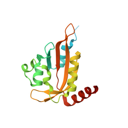
Crystal structures of Aureochrome1 LOV suggest new design strategies for optogenetics.
Mitra, D., Yang, X., Moffat, K.(2012) Structure 20: 698-706
- PubMed: 22483116
- DOI: https://doi.org/10.1016/j.str.2012.02.016
- Primary Citation of Related Structures:
3UE6, 3ULF - PubMed Abstract:
Aureochrome1, a signaling photoreceptor from a eukaryotic photosynthetic stramenopile, confers blue-light-regulated DNA binding on the organism. Its topology, in which a C-terminal LOV sensor domain is linked to an N-terminal DNA-binding bZIP effector domain, contrasts with the reverse sensor-effector topology in most other known LOV-photoreceptors. How, then, is signal transmitted in Aureochrome1? The dark- and light-state crystal structures of Aureochrome1 LOV domain (AuLOV) show that its helical N- and C-terminal flanking regions are packed against the external surface of the core ¦Â sheet, opposite to the FMN chromophore on the internal surface. Light-induced conformational changes occur in the quaternary structure of the AuLOV dimer and in Phe298 of the H¦Â strand in the core. The properties of AuLOV extend the applicability of LOV domains as versatile design modules that permit fusion to effector domains via either the N- or C-termini to confer blue-light sensitivity.
Organizational Affiliation:
Department of Biochemistry and Molecular Biology, University of Chicago, 929 E 57th Street, Chicago, IL 60637, USA.


