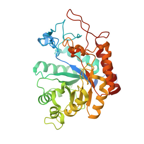
Structural and Mechanistic Insight Into N-Glycan Processing by Endo-Alpha-Mannosidase.
Thompson, A.J., Williams, R.J., Hakki, Z., Alonzi, D.S., Wennekes, T., Gloster, T.M., Songsrirote, K., Thomas-Oates, J.E., Wrodnigg, T.M., Spreitz, J., Stutz, A.E., Butters, T.D., Williams, S.J., Davies, G.J.(2012) Proc Natl Acad Sci U S A 109: 781
- PubMed: 22219371
- DOI: https://doi.org/10.1073/pnas.1111482109
- Primary Citation of Related Structures:
4ACY, 4ACZ, 4AD0, 4AD1, 4AD2, 4AD3, 4AD4, 4AD5 - PubMed Abstract:
N-linked glycans play key roles in protein folding, stability, and function. Biosynthetic modification of N-linked glycans, within the endoplasmic reticulum, features sequential trimming and readornment steps. One unusual enzyme, endo-¦Á-mannosidase, cleaves mannoside linkages internally within an N-linked glycan chain, short circuiting the classical N-glycan biosynthetic pathway. Here, using two bacterial orthologs, we present the first structural and mechanistic dissection of endo-¦Á-mannosidase. Structures solved at resolutions 1.7-2.1 ? reveal a (¦Â/¦Á)(8) barrel fold in which the catalytic center is present in a long substrate-binding groove, consistent with cleavage within the N-glycan chain. Enzymatic cleavage of authentic Glc(1/3)Man(9)GlcNAc(2) yields Glc(1/3)-Man. Using the bespoke substrate ¦Á-Glc-1,3-¦Á-Man fluoride, the enzyme was shown to act with retention of anomeric configuration. Complexes with the established endo-¦Á-mannosidase inhibitor ¦Á-Glc-1,3-deoxymannonojirimycin and a newly developed inhibitor, ¦Á-Glc-1,3-isofagomine, and with the reducing-end product ¦Á-1,2-mannobiose structurally define the -2 to +2 subsites of the enzyme. These structural and mechanistic data provide a foundation upon which to develop new enzyme inhibitors targeting the hijacking of N-glycan synthesis in viral disease and cancer.
Organizational Affiliation:
Department of Chemistry, University of York, Heslington, York YO10 5DD, United Kingdom.


