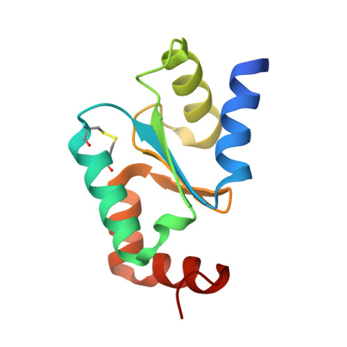
Atomic resolution crystal structure of glutaredoxin 1 from Plasmodium falciparum and comparison with other glutaredoxins.
Yogavel, M., Tripathi, T., Gupta, A., Banday, M.M., Rahlfs, S., Becker, K., Belrhali, H., Sharma, A.(2014) Acta Crystallogr D Biol Crystallogr 70: 91-100
- PubMed: 24419382
- DOI: https://doi.org/10.1107/S1399004713025285
- Primary Citation of Related Structures:
4HJM, 4MZB, 4MZC - PubMed Abstract:
Glutaredoxins (Grxs) are redox proteins that use glutathione ((¦Ă)Glu-Cys-Gly; GSH) as a cofactor. Plasmodium falciparum has one classic dithiol (CXXC) glutaredoxin (glutaredoxin 1; PfGrx1) and three monothiol (CXXS) Grx-like proteins (GLPs), which have five residue insertions prior to the active-site Cys. Here, the crystal structure of PfGrx1 has been determined by the sulfur single-wavelength anomalous diffraction (S-SAD) method utilizing intrinsic protein and solvent S atoms. Several residues were modelled with alternate conformations, and an alternate position was refined for the active-site Cys29 owing to radiation damage. The GSH-binding site is occupied by water polygons and buffer molecules. Structural comparison of PfGrx1 with other Grxs and Grx-like proteins revealed that the GSH-binding motifs (CXXC/CXXS, TVP, CDD, Lys26 and Gln/Arg63) are structurally conserved. Both the monothiol and dithiol Grxs possess three conserved water molecules; two of these were located in the GSH-binding site. PfGrx1 has several polar and charged amino-acid substitutions that provide structurally important additional hydrogen bonds and salt bridges missing in other Grxs.
Organizational Affiliation:
Structural and Computational Biology Group, International Centre for Genetic Engineering and Biotechnology (ICGEB), Aruna Asaf Ali Road, New Delhi 110 067, India.


