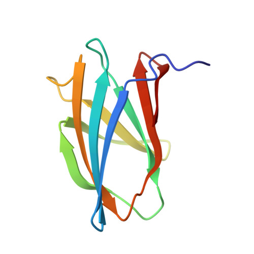
Crystal structures of chitin binding domains of chitinase from Thermococcus kodakarensis KOD1
Hanazono, Y., Takeda, K., Niwa, S., Hibi, M., Takahashi, N., Kanai, T., Atomi, H., Miki, K.(2016) FEBS Lett 590: 298-304
- PubMed: 26823175
- DOI: https://doi.org/10.1002/1873-3468.12055
- Primary Citation of Related Structures:
5DHD, 5DHE - PubMed Abstract:
Chitinase from T.?kodakarensis (TkChiA) catalyzes the hydrolysis of chitin. The enzyme consists of two catalytic and three binding domains (ChBD1, ChBD2 and ChBD3). ChBD2 and ChBD3 can bind to not only chitin but also cellulose. In both domains, the intervals of the side chains of the three tryptophan residues, which are located on the molecular surface, correspond to twice the length of the lattice of the chitin. A binding model with crystalline chitin implies that the tryptophan residues and a glutamate residue interact with the hexose ring by CH-¦Đ interactions and the amide group by a hydrogen bond, respectively.
Organizational Affiliation:
Department of Chemistry, Graduate School of Science, Kyoto University, Sakyo-ku, Japan.


