


News
Explore Sequence-Structure Relationships
08/25
Protein Feature View visually maps a PDB chain (or "instance") to corresponding sequences from UniProtKB and annotations from external resources in different "tracks" to enable explorations of structural and biological features.
This tool integrates
- Secondary structure, angle/distance outliers, protein-ligand binding sites or disulfide bridges (from PDB data)
- Structural domains (from CATH and SCOPe)
- Biochemical and biomedical features (from UniProtKB)
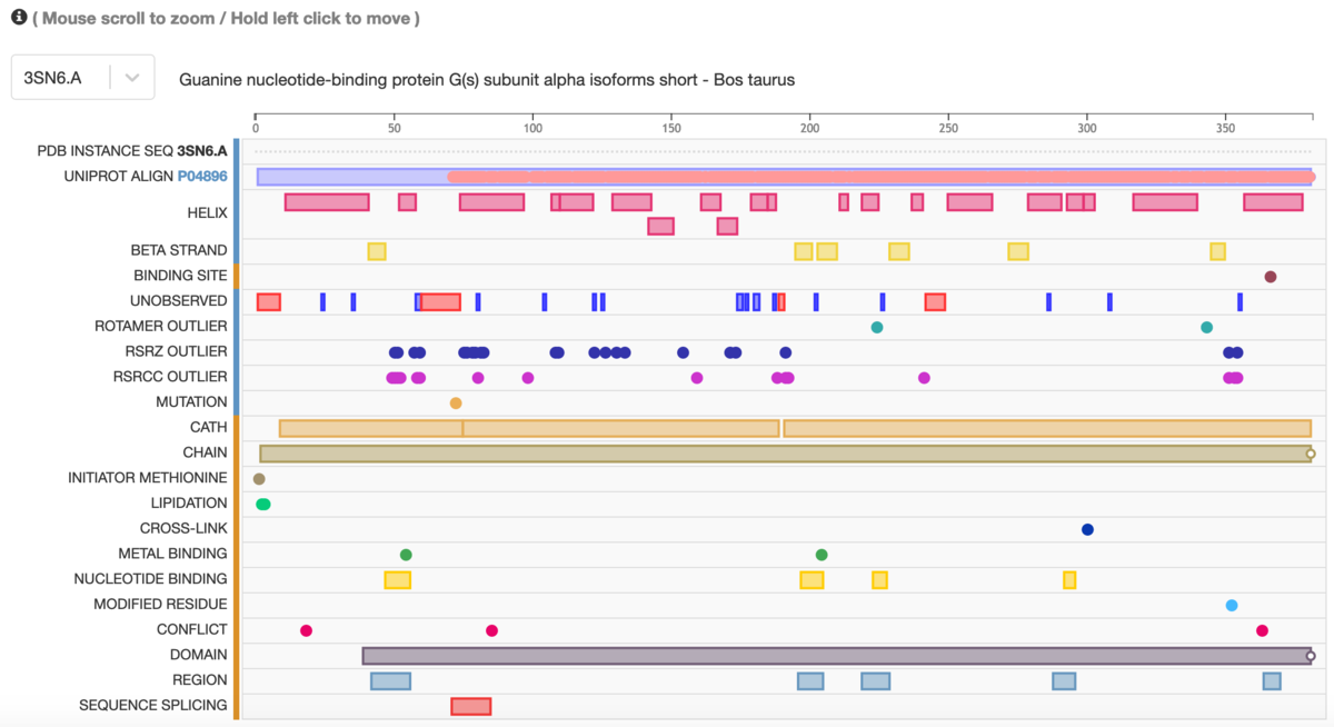 Protein Feature View for 3sn6. Zoom in with the mouse or two fingers for a granular view that includes amino acids; click to highlight specific areas; hold and click to move left/right
The Macromolecules section on a Structure Summary Page provides a truncated Protein Feature View. Selecting the Sequence tab launches the expanded view for the structure.

Protein Feature View also maps a UniProt sequence to all corresponding structures in the PDB. Access this feature from the Macromolecules section on a Structure Summary Page or enter the UniProt ID in the URL rcsb.org/uniprot/
Visit the Help Documentation for complete details.


